Division of Radiation Oncology
Radiation Oncology at University of Iowa Health Care, formerly University of Hospitals and Clinics (UIHC), did not become an independent department until 2001. Howard LaTourette, MD was appointed the leader of the Division of Radiation Oncology by Eugene Van Epps, MD — then chair of the Department of Radiology — around 1960. Originally located in General Hospital just north of the fountain entrance, it was shaped into a cohesive division by the recruitment of David Hussey, MD by Edmund (Tony) Franken, MD who succeeded Van Epps as the chair of the Department of Radiology in 1973. Hussey was instrumental in establishing a formal residency program including the practice of studying sub-specialty areas from faculty. He inherited a staff of five physicians, two physicists and a handful of therapists.
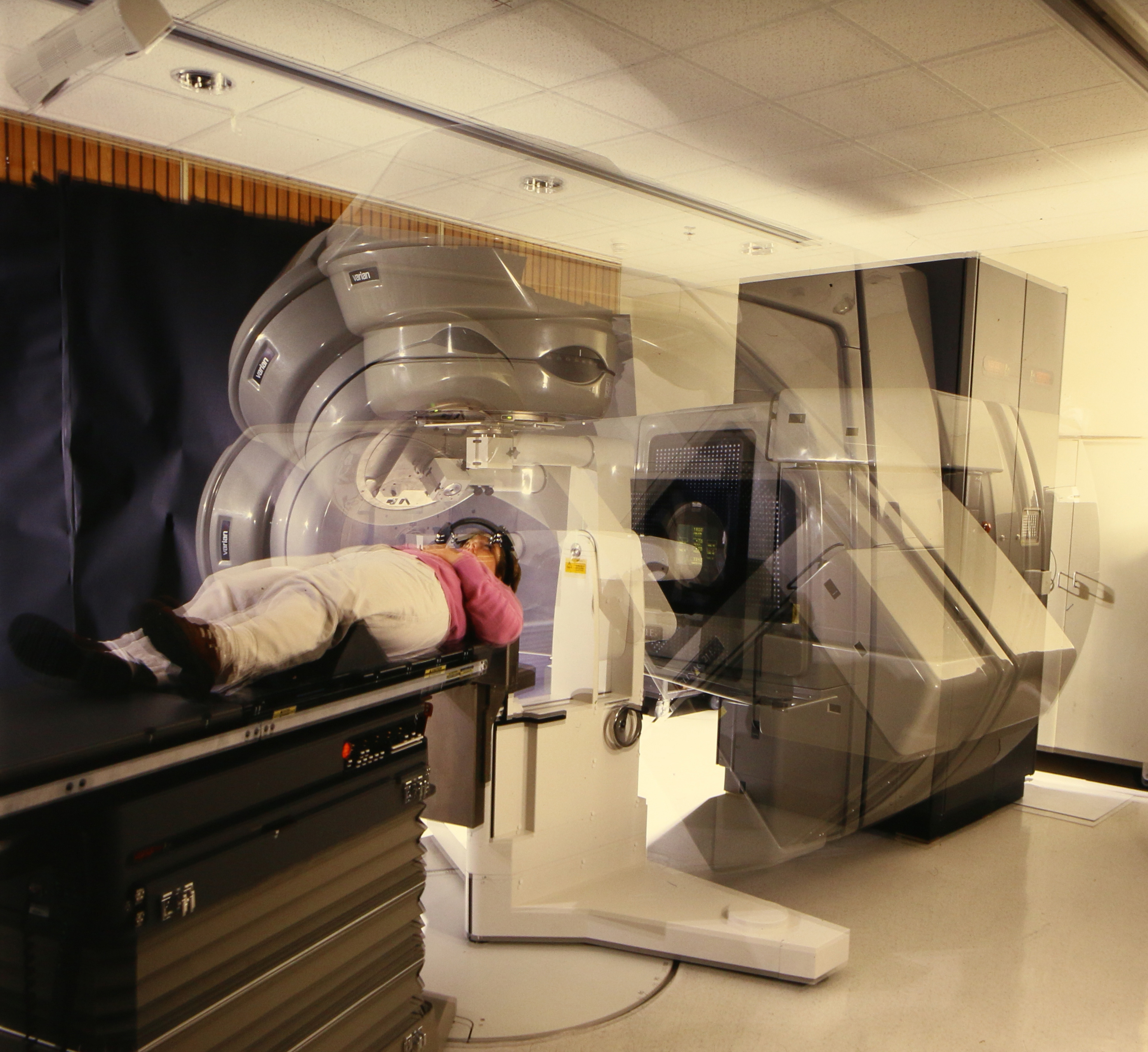
Treatment was conducted in three rooms using a cobalt machine, a single energy Varian Clinac 4 and a Varian Clinac 18. The Clinac 18 was immediately replaced with a Clinac 2500, a dual energy machine of 24 and 6MV plus electrons, thus a far more useful tool for delivering radiation therapy than the single energy devices previously used.
None of the rooms housing the type of linear accelerators installed were purpose built to accommodate them. This presented two problems: inadequate shielding and lack of space.
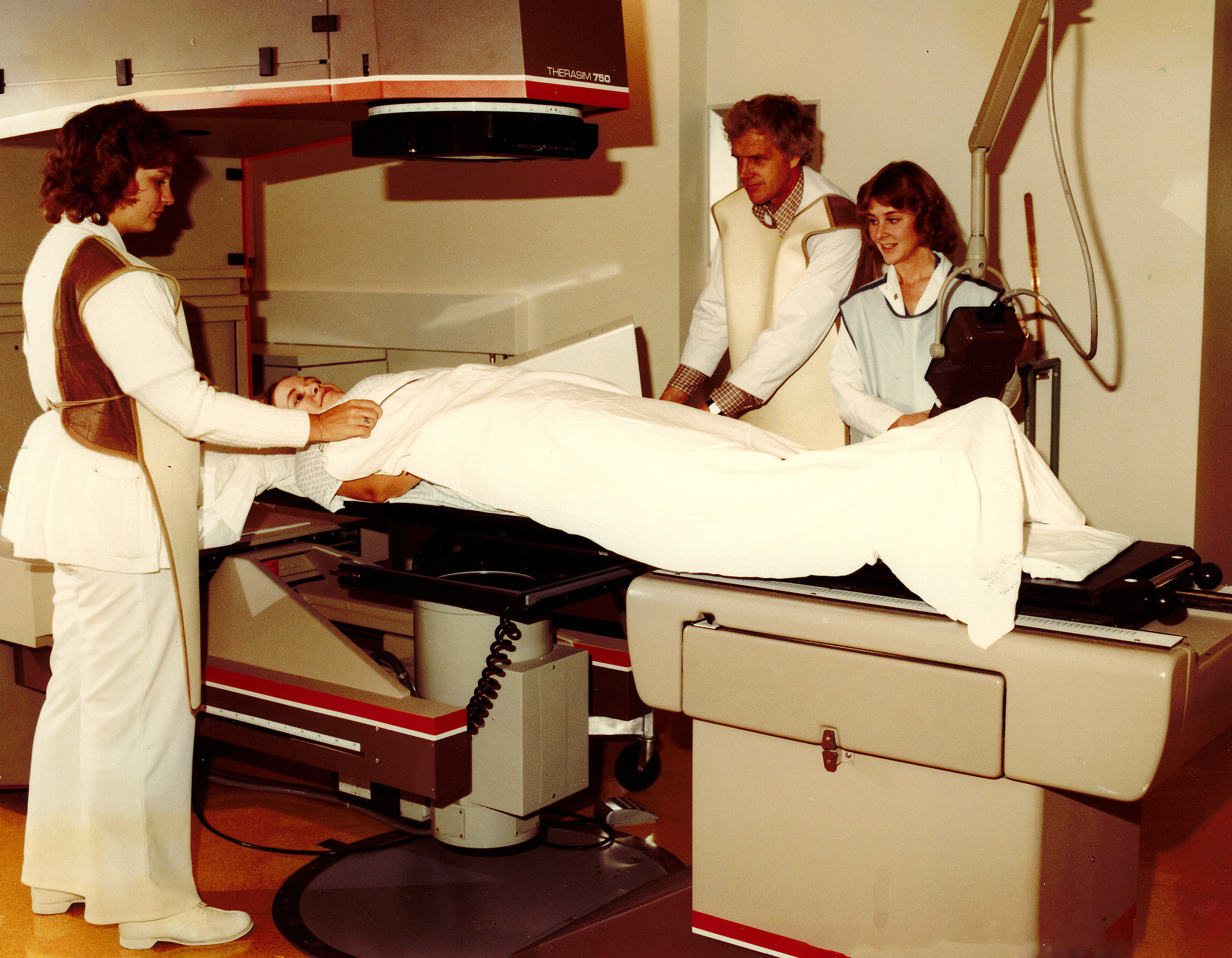
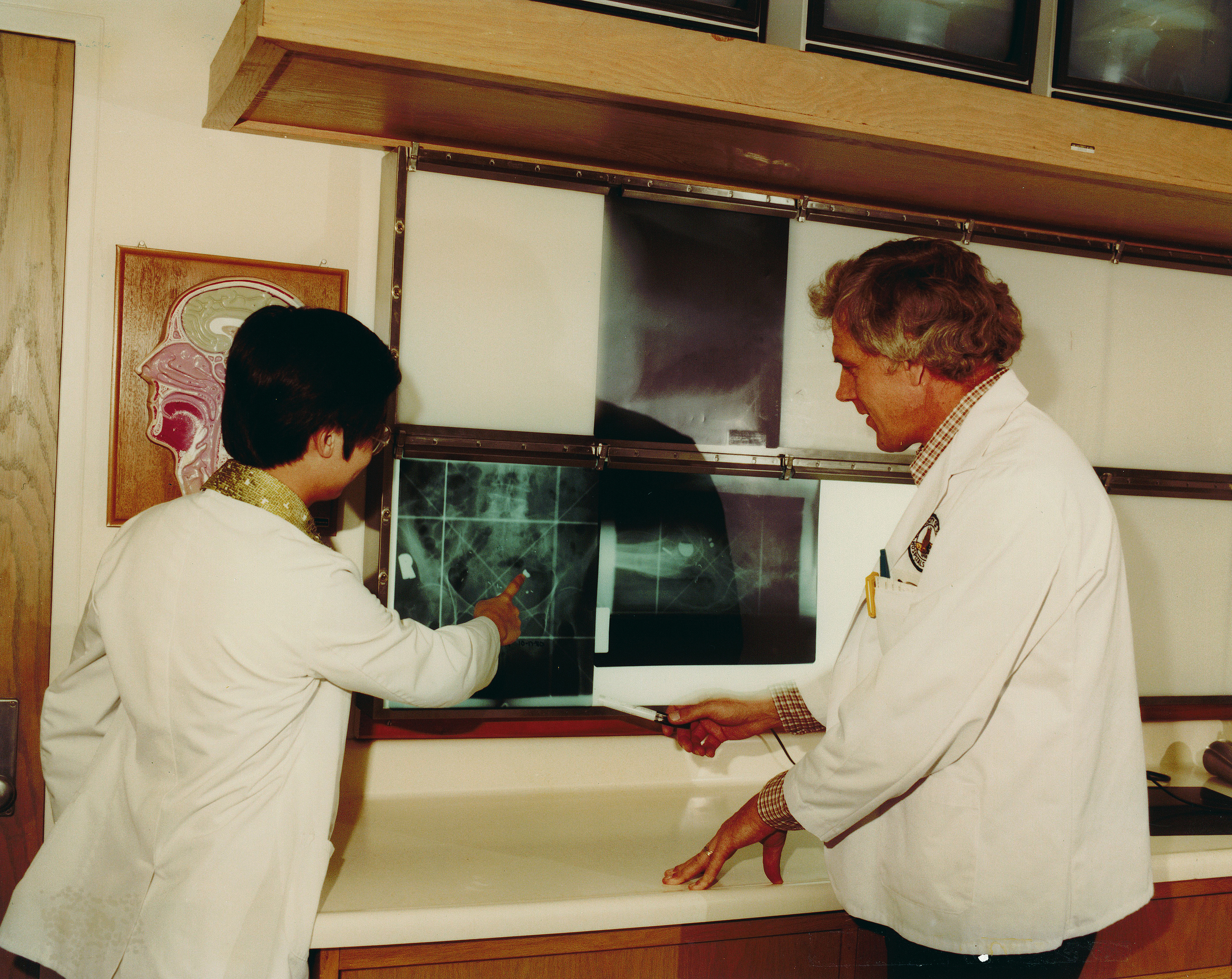
Treatment techniques of the time seem rudimentary; patients were placed in the treatment position and the simulator rotated to match the beam angles to be treated. This produced a 17 x 14 inch slice of film that would be used by the radiation oncologist to mark the area to be treated using a wax pencil or china marker. Patterns matching the area to be treated were cut on Styrofoam then used to make the Cerrobend blocks. Each treatment would require two to four custom blocks that were stored in cubbies in the treatment room.
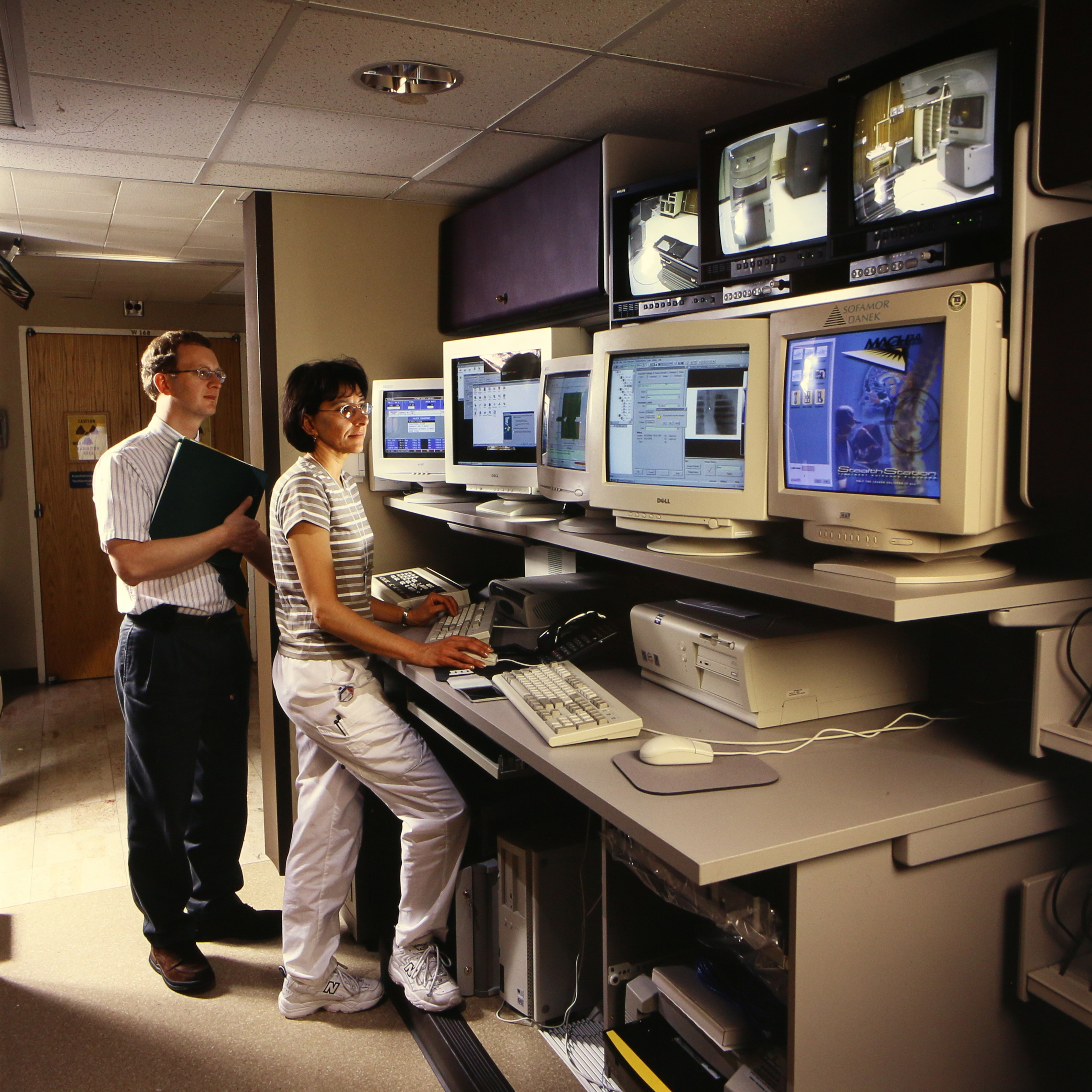
During the mid-1990’s Radiation Oncology at UIHC become one of the first clinics in the nation to use a dedicated computed tomography machine for simulation. It was comparatively slow though, taking about 60 seconds to reconstruct an image; 15 minutes to reconstruct several slices. By comparison, our current Siemens Biograph Vision reconstructs over 200 slices in one minute. During the late 1990’s, a Peacock device was attached to the Clinac 18 providing the department with its first Intensity Modulated Radiation Therapy (IMRT) capability. IMRT was first used in 1998, and calculations were performed on a computer system named “Nomos." Hussey left the University of Iowa in 1999, took a sabbatical year in Tucson, then accepted a position at the University of Texas at San Antonio in 2000.
Early Beginnings
Building CEIGRT
From Division to Department
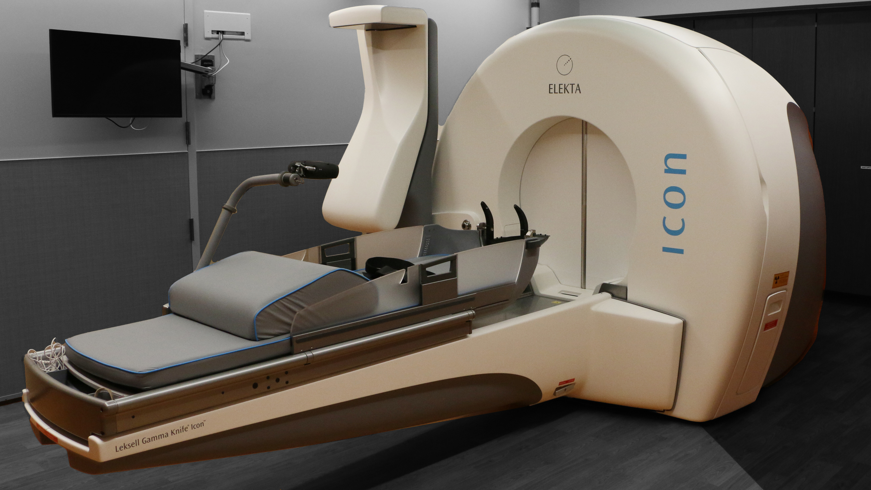
Following the departure of David Hussey MD, University of Iowa Health Care recruited John M. Buatti, MD from the University of Florida who started on October 1, 1999. He introduced a stereotactic radiosurgery (SRS) technique for linear accelerators, established peer pay-parity for the radiation therapists and moved the department towards independence from radiology. On July 1, 2001, we became the Department of Radiation Oncology allowing greater freedom to expand as needed.
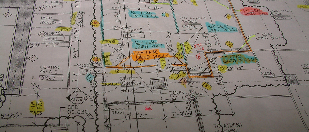
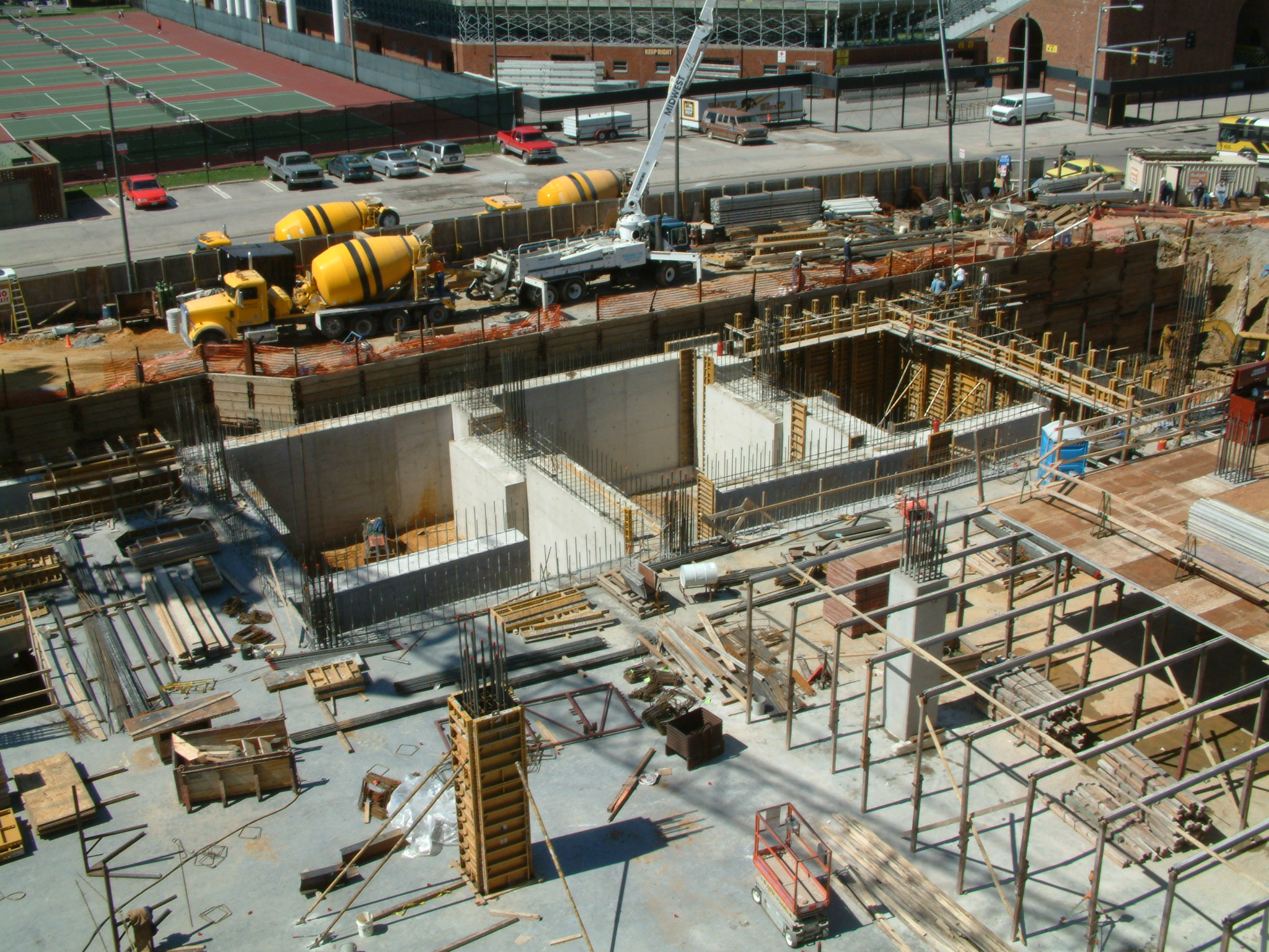
In order to meet the expected increase in patient numbers and provide a comfortable, flexible space in which to treat them, work began on building the Center of Excellence in Image Guided Radiation Therapy (CEIGRT). Careful consideration was given to the construction of the vaults housing the treatment devices. Particular attention was paid to providing enough room for any potential growth in size and sophistication while still having sufficient room to keep patients and staff comfortable.
Construction began in 2002 to the north of Pomerantz Family Pavilion. The site was noted for having the deepest hole dug in Iowa City at the time. Construction and commissioning of the devices was completed by May of 2005 and the clinic opened in June 2005.
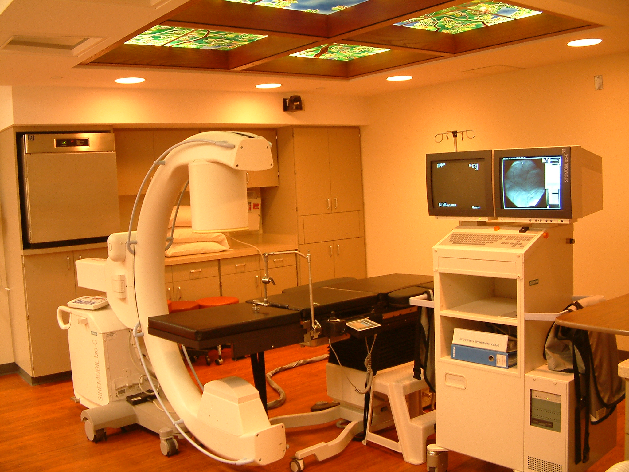
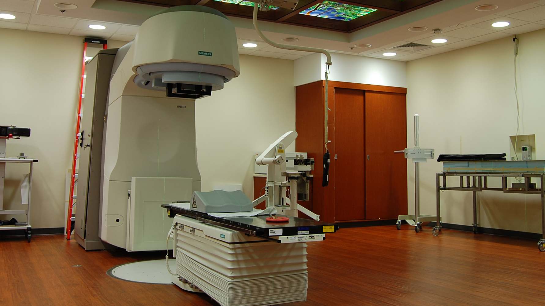
The 40,000 square feet CEIGRT was equipped entirely with Siemens devices for imaging, simulation and treatment, establishing the world’s first completely image and optic-guided stereotactic radiosurgery system. Having a single vendor provide all the imaging and treatment devices gave the ability to simulate patients on the treatment devices as well as having seamless image integration throughout the entire treatment process. Soft tissue imaging for genitourinary and chest cancers used a 3T MRI, providing image clarity previously unknown. The department was one of the first to offer high-dose rate brachytherapy treatments in the region and attracted much interest from peer groups wishing to implement a similar program.
Growth and Expansion
Since 2005 the department has grown and expanded consistently each year. This period of growth was characterized by research supported by securing major grants in imaging (Quantitative Imaging to Assess Response in Cancer Therapy Trials) and ascorbate research (Exploiting Redox Metabolism Using Pharmacological Ascorbate for Cancer Therapy). These projects yielded therapies and techniques that directly improve patient care. An innovative medical physics division pioneered 3T MRI imaging coupled with titanium tandem & ovoid brachytherapy, developed a dynamic rotating shield brachytherapy applicator and dynamic collimation system for proton therapy.
MBA, MD, PhD
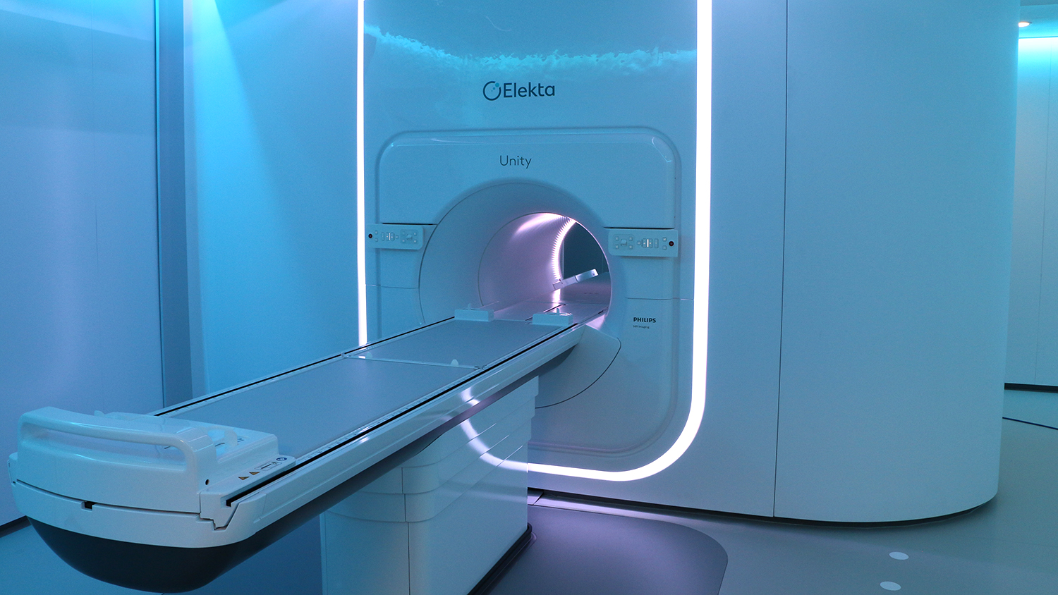
Our department was the third in the nation to commission an Elekta Unity magnetic resonance linear accelerator (MR-Linac). It provides imaging during treatment allowing the treatment team to precisely target the tumor in real time and is especially beneficial for seeing soft-tissue tumors of the lower abdomen.
In the near future the Iowa FLASH laboratory will commission the Radia Beam Flex-9 FLASH irradiator, the first of its kind in the US and vital to universal FLASH development and research.
Bryan G. Allen, MBA, MD, PhD succeeded John M. Buatti, MD as Chairman and DEO of the department on July 1, 2023, bringing his extensive expertise in translational research to the position.
Innovative Research and Treatment