Available Equipment
The Department of Radiation Oncology resides in a 40,000 square foot facility. This facility is equipped with three Elekta Versa HD linear accelerators, an Elekta Unity MR-Linac, and one Elekta Gamma Knife Icon stereotactic radiosurgery system.
- 48 servers consisting of a mix of Physical and Virtual servers, running various OS systems like Windows Server 2016-2022, Linux CentOS, Ubuntu, and Unix.
- Our PACS has 25TB of image storage space
- 1 prostate workstation
- 2 Oncentra Brachy Workstations (1 server and 1 client)
- 3 Siemens Workstations
- 1 Fuji Film server for maintaining filming without paper
- In our secondary Clinic, we have 30 workstations and 3 servers running Windows Server OS
- 200 workstations in the department
Biograph Vision PET/CT
- The Siemens Biograph Vision device incorporates a 3.2 x 3.2 x 20-mm crystal element that individually selects and delivers high 48-mm3 isotropic spatial resolution; 100% coverage of the crystal area with SiPM sensors providing a timing resolution of 214 picoseconds, with 3.9 times higher effective sensitivity for faster scans and lower dose.
- Four Dimensional Computed Tomography (4DCT) and Positron Emission Tomography (4DPET) allows the patient internal movement due to respiration to be determined.
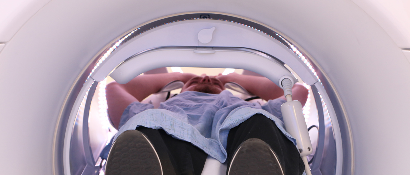
Siemens Magnetom Vida MRI
The Siemens Magnetom Vida provides enhanced MR-imaging capability and efficiency, including receiver technology of 64 channels, gradient coil strength to 60 mT/m per axis, and a bore of 70 cm with a large field-of-view of 55 cm guaranteed homogeneity on a 50 cm field-of-view, compressed sensing acquisition and reconstruction, respiratory triggered imaging, and four- dimensional (4D) MRI. The radiofrequency (RF) coils include the CP-head, 64 channel head / neck array, 18 channel body array, along with large and small flexible array coils.
Linear Accelerators
The three Elekta Versa HD linear accelerators have 6, 10, and 18 MV x-ray beams and 6-15 MeV electron beams, 160 leaf multi-leaf collimators (MLCs), electronic portal imaging, and kilovoltage computed tomography (kV-CBCT) imaging for daily patient positioning in 3-dimensions and 4-dimensions. The Versa HDs can treat patients using respiratory gating and the Varian Optical Guidance Platform (OGP), which enables real-time patient positioning and motion correction under infrared camera guidance for brain cancer patients. Two of the Versa HDs are capable of delivering total body irradiation and one is capable of total skin electron therapy. CIVCO Protura six degree-of-freedom couch tops are available for patient position adjustments in nearly real-time. The Versa HD accelerators are capable of volumetric modulated arc therapy (VMAT), which delivers highly conformal radiation therapy dose distributions rapidly, in 5-10 minutes.
The Elekta Unity MR-Linac
This device has a single photon energy radiation beam that delivers radiation through the wall of a wide bore (70 cm) lengthened 1.5 Tesla Philips MRI scanner. In addition to being able to provide exquisite soft tissue image contrast for image guided radiotherapy, the system is the first to have the capability to "see what you treat" during radiation therapy and respond during treatment based on the imaging information. The Elekta Unity MR-Linac includes specialized software that provides adaptive radiation therapy capability based on up-to-date information on the patient's internal anatomy at the time of treatment.
Gamma Knife Icon Stereotactic Radiosurgery System
The Gamma Knife Icon is capable of delivering both frame-based and frameless stereotactic radiosurgery. The system has 192 separate 60Co radiation sources distributed over eight sectors oriented in a cone shape around the patient's head, and at a given time during treatment each sector can be configured to deliver 24 individual, focused, gamma ray beams with circular field sizes of 4 mm, 8 mm, 16 mm, or no beams. When treating patients with a stereotactic frame, the system is capable of 0.3 mm accuracy. A kilovoltage cone beam CT gantry is mounted to the system, providing imaging capability for frameless stereotactic radiosurgery.